Archiving DICOM images
Backup for data recovery after hardware failure
Database for queries DICOM query/retrieve ( FIND/MOVE SCU/SCP) from remote DICOM workstation
Routing Functions
Support for DICOM worklist and Support for DICOM MPPS server.
Support for HL7 – ADT, ORM, sending reports
Access control for different levels of users with userid/password
Teleradiology ready
System management and archiving of written medical report attached to each examination
Ability to load in the local database
Browser based workstations is capable of displaying images for diagnostic purpose on 1 high resolution monitor and has DICOM and HL7 interface to do all the above functions. Modality support for CT, MR, US and image mgmt features for the same
Diaphragm function
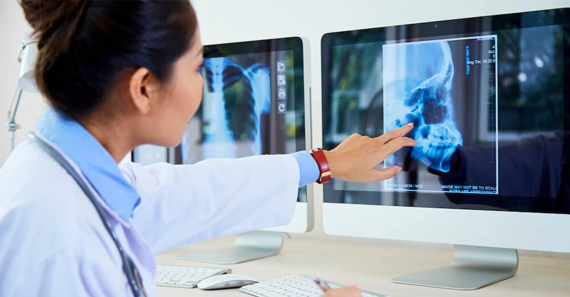
Image processing, - all features windowing, zoom, magnifiying glass, annotations, density calculation(CT) and measures ( area, distance, angle …)
Pagination and printing images on standard windows printer and imager (DICOM Print)
Pre defined filters and user defined filters
Burning CD/DVD images of all modalities with a CD viewer for distribution.
Flips, rotates, inverts the images
Open or close list of images, select the previous and next study, resets the original images, adjusts the brightness, the color, the contrast and performs all functions
The, html5 zero-footprint DICOM Viewer has an extensive radiology tool set, which includes regular features such as
Window. Pan. Zoom, Scroll. Rotate/Flip. Magnifier.
The DICOM Viewer advanced features and radiology measurements are:
Line. Angle. Cobb angle. Polyline,Area. Ellipse. Volume. CTR. (Measure a cardiothoracic ratio to estimate a heart size.)
Foot longitudinal arch, Spine labelling ,ROI. Flexpoly, Pencil. Arrow. Text
Intensity. Measurement to measure Hounsfield units at a specific point of a CT study;
Show angles. Calibration line. Delete annotation. Save annotation.
The web DICOM Viewer has an advanced layout features for Layout, Thumbnails, Multi-image. Key Objects storing and Hanging protocols
The DICOM Viewer also has the image manipulation features such as Reference lines, Orientation labels, Link Series, Cine Mode and crosshair.
The DICOM Viewer also has specific features including multi-frame and video support:
ECG support. Electrocardiography study support;
PDF support. Support for PDF modality;
SR support. Support for SR modality;
Key Objects (KO) support. Possibility to mark instances as KO and save them;
Fusion. PET-CT Fusion allows combining the series of PET and CT types, thus linking the sites of radioactive drug concentrations with the patient's anatomical structure;
Color channels. Highlight a color component or a combination of them;
MPR. Multi-planar reconstruction with auto rotate Coronal or Sagittal projections
The 3D DICOM Viewer option has the standard tools (pan, zoom, rotation rendering settings presets) and advanced MPR features:
Oblique MPR.
MIP
MinIP
AVG (AvIP, AIP)
Features of 2D. 3D rendering
MPR / MIP comparison